Definition of Human Heart
Human heart or heart is that part of the body which is mainly known for pumping blood in our body with the help of blood system.
In addition, it reaches oxygen and nutrients to the tissues and works to remove carbon dioxide and other dissolved things. Our heart beats an average of 72 times a minute.Our body parts need oxygen supply to function. If our heart does not flow smoothly to the body parts, it can prove to be dangerous. Our heart is found in the middle of both lungs, in the middle part of the chest.
Structure of Human Heart
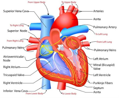
The heart is a muscle whose size is slightly larger than the fist. Like other muscles of the body, it keeps shrinking and expanding. Whenever this organ is extended, it extends with full force, while the other organs expand depending on the sequence. Whenever blood is pulsed from the heart or pumped to other organs, this reaction is called a cardiac cycle, which appears 72 times a minute.Heart weight varies from 280 to 340 grams in men and 230 to 240 grams in women. This organ is divided into four chambers - the two chambers at the top are called the arteria and the two chambers at the bottom are called the ventricle. The right side arterium and ventricle together form the right heart, in the same way the left heart is. The thin wall of the muscle, called the septum - divides the right and left heart into two parts.
A double-layer membrane called a pericardium acts as a shell or covering for the heart. The outer layer of the pericardium is called the parietal pericardium and the inner layer is called the serous pericardium - they contain pericardial fluid that protects the heart from the effects of contractions and motion of the lungs and diaphragm.The outer layer of the heart has three layers. The outer layer is called the epicardium. The middle layer is called the myocardium - inside these are found the muscles that keep shrinking. The inner lining or anocardium is exposed to blood.
The arteries and ventricle are connected by the artrioventricular valves, which consist of the tricuspid valve and the mitral valve. Pulmonary semilunar valves work to separate the right ventricle and the pulmonary artery. The aortic ventricle functions to separate the left ventricle from the aorta. With the help of valve heartstrings, the muscles are vaccinated.
Function of Human Heart
Our heart carries blood through two paths -
Pulmonary circuit
From here, oxygen-free blood passes through the pulmonary artery to the lungs through the right ventricle and then through the pulmonary vein in the form of blood with oxygen to the left arterium.
Systematic circuit
In this part, blood including oxygen goes through the left ventricle into the aortic, then into the capillaries, from where they provide oxygen to various organs of the body.


No comments:
Post a Comment